


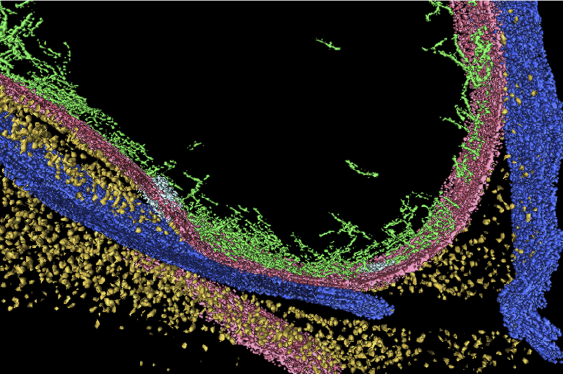
Cryo electron tomography (CET) reveals the macromolecular and structural organization within cells in a close-to-life state. It is, however, limited to thin specimens, such as peripheral areas of intact eukaryotic cells. Analysis of tissue ultrastructure requires physical thinning prior to the application of CET.
Ben-Gurion University of the Negev researcher Prof. Ohad Medalia and his colleagues have further developed and demonstrated the use of ion beams to thin samples for CET imaging of vitrified multicellular tissues. By combining sample freezing directly on EM grids with the means for introducing gold nanocrystals markers on the surface of the CFIB-milled lamellae, they were able to produce high quality tomograms of vitrified C. elegans embryos and worms.
The paper was just published online in Nature Methods.
“The method described here offers a general solution for acquiring high-resolution tomograms on thick cells and tissues in diverse imaging settings. CET can now be implemented to resolve macromolecular structures from tissues, thus bridging the gap between developmental, cellular and structural biology,” says Medalia, a member of the Department of Life Sciences and the National Institute for Biotechnology in the Desert.
Publication in Nature Methods, May 11th 2015